Craniosynostosis
A multidisciplinary approach to caring for craniosynostosis
What is craniosynostosis?
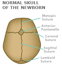
Craniosynostosis is a condition where one or more of the fibrous connections between the brain and the skull, called sutures, in a child's skull closes too early. When that occurs, it alters the skull's growth pattern.
When the sutures are open, they allow the skull to grow at the same rate as the brain. In the first few years of life this growth is very rapid.
If a suture closes too early, the skull cannot grow normally, which causes pressure on the growing brain. As the brain continues to grow, it pushes against, or expands, the areas of the skull that are not yet fused. This leads to a change in the shape of the head.
Types of craniosynostosis
There are four types of craniosynostosis:
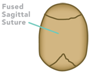
Sagittal craniosynostosis (scaphocephaly)
Sagittal craniosynostosis is the most common type. The sagittal suture runs from the baby's soft spot at the top of the head (anterior fontanelle) straight back.
When it fuses too early, a raised ridge can often be felt or even seen over this area and the soft spot may be absent or small. As the brain grows, the skull can no longer get wider, so the skull gets longer and bulges at the front and the back. A baby with sagittal craniosynostosis may seem to have a large or prominent forehead.
Read Keith's story to learn more about this condition.
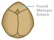
Metopic craniosynostosis (trigonocephaly)
The metopic suture runs from the baby's soft spot at the top of the head (anterior fontanelle) to the forehead. When this suture fuses too early, a raised ridge can often be felt or even seen over this area and the soft spot may be absent or small.
As the brain grows, the forehead can no longer get wider, and appears pinched, or pointed. When viewed from above, the head shape looks like a triangle. A baby with metopic craniosynostosis may seem to have their eyes close together.
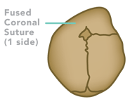
Coronal craniosynostosis
The coronal suture runs from a baby's soft spot at the top of the head (anterior fontanelle) toward the ear on both sides. When it fuses too early on one side (called unicoronal), the forehead looks flat on the affected side.
The eyebrow can appear higher and as the brain grows the forehead can appear more prominent on the unaffected side. If both sides are affected (called bicoronal), the forehead may look tall, Also, when viewed from above the shape of the head will be wide.
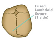
Lambdoid craniosynostosis (posterior plagiocephaly)
The lambdoid suture is found in the back of the head. When this suture fuses too early, the back of the head is flattened, and the ear may be lower on the affected side. This is the rarest type of craniosynostosis.
What causes craniosynostosis?
The exact cause of craniosynostosis is not yet clear. In about 20 percent of children with single suture craniosynostosis, a mutation or change can be identified in one of the genes. (Genes determine the characteristics or traits that pass from a parent to a child.) Craniofacial syndromes may also be related to a gene mutation.
There is scientific evidence that this premature fusion occurs when the bones of the skull are restricted in the womb, not allowing for movement of the growing brain. This may be why craniosynostosis occurs more often in twin pregnancies.
If a couple has a child with single suture craniosynostosis, the chance their next baby will also have craniosynostosis is believed to be less than 2 percent. Our center is currently investigating both the physical and underlying genetic causes of craniosynostosis.
How do you test and diagnose craniosynostosis?
If a family or pediatrician notices an infant has an unusual head shape, an unexpected change in head size (circumference) or early closure of the soft spot (anterior fontanelle), the infant should be referred to a craniofacial specialist for evaluation.
The most common reason for a baby to have an unusual head shape is not craniosynostosis, but a condition called plagiocephaly which does not involve a fused suture.
An experienced pediatric craniofacial surgeon or pediatric neurosurgeon at our craniofacial clinic can make the diagnosis by physical exam. In some cases, a medical image like a CT scan of the skull may be obtained to confirm the diagnosis and assist with the evaluation.
How is craniosynostosis treated?
Surgical treatment is recommended when craniosynostosis affects the shape of the head in a significant way. The more the shape of the head is affected, the greater the worry about the effect on the child's brain. Surgery is believed to give a child the best possible chance to develop to their full potential. It also normalizes the child's appearance. An unusual head shape can affect a child's personality, self-esteem and social interactions.
Our craniofacial specialists have two approaches to repairing craniosynostosis:
During this surgery, the surgeon removes the fused suture through one or two small cuts (incisions). The surgery does not involve active reshaping of the head. Instead, the baby wears a helmet to mold their head to a more normal shape in the months after surgery.
This may be a treatment option if a baby is six months or younger. After six months of age, a baby's head does not grow as fast and helmets are not as effective.
About three to four days after endoscopic strip surgery, the baby will be measured for a helmet. The specialist who measures and makes the helmet is known as an orthotist. Using gentle pressure, the helmet molds the baby's head as the skull heals and grows. It also keeps the suture from fusing again too soon. Babies wear the helmet for at least three months and sometimes for up to a year, typically for 23 hours a day. A baby may need a new helmet made every several months to keep up with the growth of their head.
Learn more about minimally invasive craniosynostosis treatment
The goal of this craniosynostosis surgery is to expand the amount of space in the skull to relieve pressure on the brain and create a normal head shape. A slight overcorrection of the head shape is performed to allow for future growth. The surgery is performed by our craniofacial surgeon and pediatric neurosurgeon.
Each specialist performs the part of the operation in their area of expertise and working together as a team, the surgeons are able to reduce the amount of time a child requires under anesthesia. Average surgical times at our hospital are around three hours.
Risk and benefits
Some families prefer the benefits of the minimally invasive endoscopic option, which may include smaller incisions and less scarring and bleeding. Others prefer cranial vault remodeling as helmets are not required with this technique. Our pediatric craniofacial and neurosurgeons are experienced in both procedures and will discuss the treatment options for your family. Families are given as much time as needed to feel comfortable with the treatment plan and have their questions answered.
Surgical treatment is generally performed before the child turns one year of age. Exact timing is based on which suture, or sutures, are fused and it can vary from two to 10 months of age. If all the sutures are fused, earlier treatment may be recommended. When a child is diagnosed after age one, surgery may be recommended at the time of diagnosis.