Neurosurgery craniosynostosis
Craniosynostosis

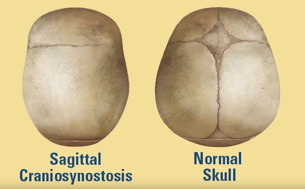
Craniosynostosis (cranio refers to head - syn means together - ostosis refers to growth of bones - thus craniosynostosis literally means head bones that grow together). This is a condition in which one or more of the fibrous gaps between infant skull bones (called cranial sutures) prematurely grow together and fuse by turning into bone (ossification). When that occurs it alters the skull's growth pattern. This happens because the skull cannot expand perpendicular to the fused suture. The shape of the head alters in compensation, as the child's brain grows.
The resulting abnormal head shape that occurs usually provides the necessary space for the growing brain, but distorts both the cranial and facial features. Sometimes the compensation does not effectively provide enough space for the growing brain. That condition is called cranial stenosis and it often results in increased intracranial pressure leading possibly to visual impairment, sleeping impairment, eating difficulties, or an impairment of mental development combined with a significant reduction in IQ.
Craniosynostosis occurs in one in 2000 births. Craniosynostosis can occur as part of a genetic syndrome in 15 to 40 percent of the patients, but it usually occurs as an isolated condition.
The operations that have been developed by pediatric neurosurgeons who work closely with plastic craniofacial surgeons can be grouped generally into two categories: cranial release operations (usually performed only on infants younger than 6 months of age) and cranial reconstruction operations (generally performed on patients older than 6 months of age).
Cranial release operations focus on removal of the offending fused suture (sometimes described as a synostectomy) together with the making of additional bone cuts that loosen the plates of bone (called "barrel stave osteotomies"). The strategy behind this approach is to release the bones sufficiently to allow the rapid brain growth, that happens naturally during an infant's first eighteen months of life, to remodel the released skull bones back to their normal shape. Sometimes the infant wears a custom-made helmet after surgery to facilitate that natural head shape remolding. This type of operation depends upon the very young age of the patient because older patients have thicker bone and slower brain growth, which prevent effect head shape remodeling in the older child.
Cranial reconstruction operations have as their primary goal to remove the abnormal bones, directly refashion them, and then replace them in such a way that it immediately restores the head fully to its correct natural shape. This can be performed at any time during the first several years of life, but is commonly performed around nine months of age. The post-operative use of a custom-made helmet is typically not required after cranial reconstruction operations.
Learn more about CHoR's Center for Craniofacial Care.
Learn more about minimally invasive craniosynostosis surgery at CHoR.
